
SciELO - Brasil - Identifying and understanding optical coherence tomography artifacts that may be confused with glaucoma Identifying and understanding optical coherence tomography artifacts that may be confused with glaucoma
Tracking laser tomography
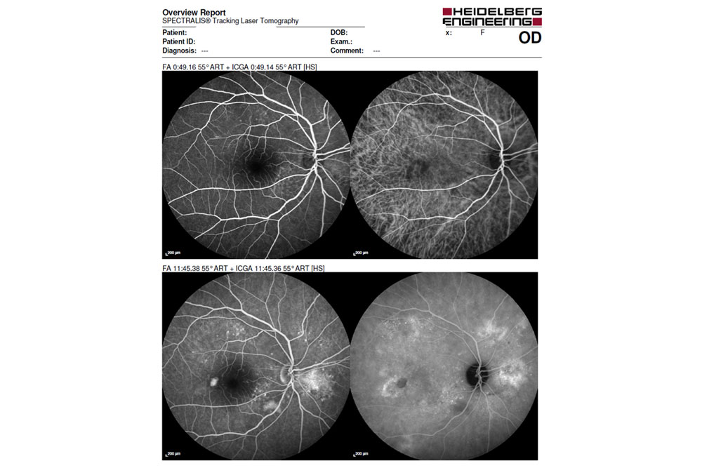
Tracking Laser Tomography. Irreversible bilateral macula edema was... | Download Scientific Diagram

Retinal vascular impairment in Wolfram syndrome: an optical coherence tomography angiography study | Scientific Reports

What OCT Tells Us About Progression

Multi-modal and multi-scale clinical retinal imaging system with pupil and retinal tracking | Scientific Reports

is a SPECTRALIS printout with the upper part showing the ONH analysis,... | Download Scientific Diagram

Artifacts and Anatomical Variations in Optical Coherence Tomography | SpringerLink

Laser Tomography - an overview | ScienceDirect Topics

Wide-field optical coherence tomography based microangiography for retinal imaging | Scientific Reports

OCT and Glaucoma: Case Review | SpringerLink

The use of Confocal Scanning Laser Tomography in the Evaluation of Progression in Glaucoma | IntechOpen

Optical Coherence Tomography - EyeWiki

Optical coherence tomography in pediatric patients: a clinical review - Bradley S. Gundlach, Irena Tsui, 2020
Tracking laser tomography

3 Optical Coherence Tomography of the Optic Nerve | Ento Key

Diagnostic Imaging Devices I OCT | Heidelberg Engineering

The use of Confocal Scanning Laser Tomography in the Evaluation of Progression in Glaucoma | IntechOpen

Scanning laser ophthalmoscopy - Wikipedia

The Anatomy of an OCT Scan

Laser Tomography - an overview | ScienceDirect Topics
; "Angiography")
Angiography

Ocular Coherence Tomography - Primary Eye Care
 of... | Download Scientific Diagram")
Optical coherence tomography/scanning laser ophthalmoscopy (OCT/SLO) of... | Download Scientific Diagram

Comparison between confocal scanning laser tomography, scanning laser polarimetry and optical coherence tomography on the ability to detect localised retinal nerve fibre layer defects in glaucoma patients | British Journal of Ophthalmology
Comparison of Optical Coherence Tomography Measurement Reproducibility between Children and Adults | PLOS ONE
